World Radiation Day - Today (November 8, 1895) is the day Willem Rontgen the first Nobel Prize of Physics discovered in X-rays.

International Day of Radiology is
a special day celebrated every year to bring the excellence of medical
morphology to the public in today's modern medical field. It is celebrated on
November 8, the day X-rays were discovered. The European Union for Radiation
and the American College of Radiation co-founded the day in 2012. Wilhelm
Conrad Rontgen was a German physicist at the University of Wurzburg on November
8, 1895, who discovered the area of waves called X-rays (X-rays) today in the
series of electromagnetic radiation. He was awarded the first Nobel Prize in
Physics in 1901 for this discovery.


Willem Rontgen was born on March
27, 1845, in Lennep, Bavaria, Germany, the only son of Friedrich Conrad Rontgen,
a textile merchant and businessman. In 1865, he studied engineering at the
Federal Polytechnic College in Zurich. He received his doctorate in 1869 from
the University of Zurich. Studies were underway in several laboratories on how
electrification occurs in low-pressure gases. Rontgen also included himself in
the review. 1895 While studying the various external effects of vacuum tube
equipment, he discovered that the barium platinosinide-coated sheet near the
negative rays formed glowed. Following that he did some more experiments in the
darkroom and realized that this glow was due to an invisible type of
radiation. However, he named it X-rays because of their unknown properties. And
then the name stuck. Two weeks later he had his wife's hand photographed for
the first time.

Rontgen published an original
article entitled A New Type of Rays on December 28, 1895. On January 5, 1896,
an Austrian newspaper published Rontgen's discovery of a new type of radiation.
He published three research papers on X-rays from 1895 to 1897. Today Rontgen
is known as the father of radiation medical testing. Rontgen never claimed
patents for his inventions. He also donated the Nobel Prize to the University
of Wurzburg.

X-rays (X-rays) are the most
powerful rays. Permeable to metals such as iron. Their wavelengths range from
10 nanometers to 0.01 nanometers. These rays are not affected by
electromagnetic fields. X-rays came into use two months after their discovery.
Radiation was used to diagnose and treat a fracture at Homsphere Hospital. Not
only does it help to penetrate the human body, but it also helps to test without
opening boxes on airfields. These rays go in a straight line. This is why they
are used to taking radiographs in diagnostic radiology.

X-rays produce biological effects. This is one of the reasons why they are used in cancer medicine. This method is called radiation therapy. These rays cause fluorescence when dropped on certain materials, such as cadmium sulfide (CdS) and sodium sulfide (ZnCdS). This feature helped to detect X-rays. It is also used in fluorescent screens and intensifying screens. They are widely used in crystallography and industry. X-rays travel at the same velocity as normal light waves. All the properties of light waves apply to this.

There is no global consensus on distinguishing between X-rays and gamma rays. They differentiate between these radiations by keeping their source. X-rays are emitted and emitted from electrons. But gamma rays are emitted from the nucleus. There are problems with this definition because it is possible to generate this high energy through other processes and sometimes it is not known how it is generated. A common alternative is to differentiate X-ray and coma radiation based on wavelength. Radiation with a wavelength of 10−11 m is called Gamma.

X-rays are high-powered
electromagnetic rays. Their ionization energy is much higher than that of
ultraviolet rays. They, therefore, have the power to ionize materials and break
chemical bonds. Because of this, X-rays are considered extremely dangerous to
organisms. Prolonged handling of sensitive X-rays without proper protection can
lead to disruption of the order of DNA molecules and cancer. High-power X-rays
can ionize and destroy cancer cells. However, X-rays of healthy cells should be
avoided. They are only used if the benefits of using X-rays in medicine
outweigh the risks.

Strong X-rays can penetrate
objects. This property is used in the medical field to take X-rays. The same
technology is used during security checks at airports. The light emitted by
X-rays is much shorter wavelengths than UV rays. So microscopes that use X-rays
are much clearer than light microscopes.

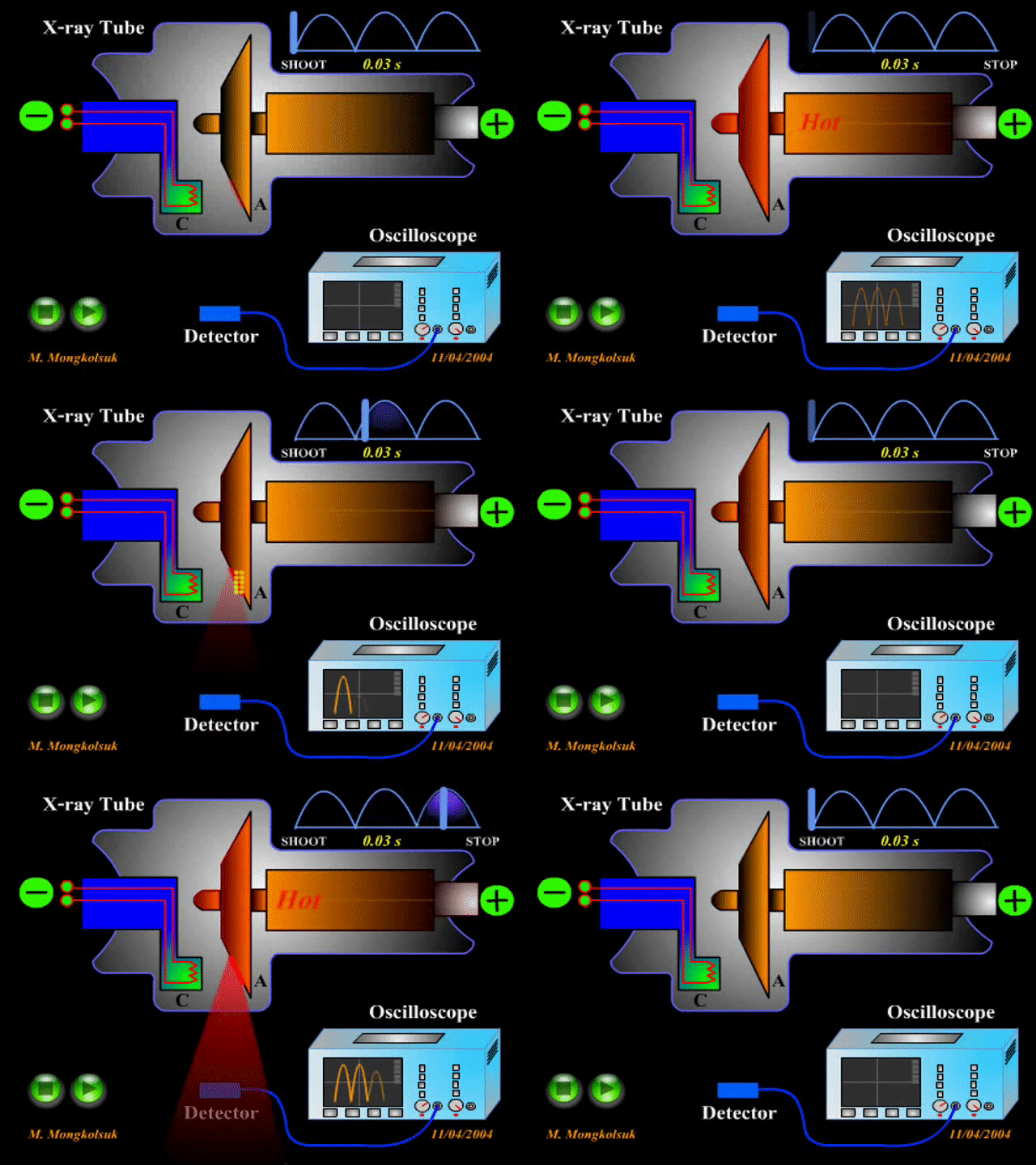
Although X-rays are emitted from
electrons, they can also be produced from a vacuum tube X-ray tube, which
accelerates the electrons emitted by the thermal electrode using high voltage.
Electrons are injected into the low-pressure tube from the opposite (cathode)
mouth of the radiant tube. Electrons travelling at high speeds collide with the
metal target anode (anode) to form X-rays. Tungsten or slightly rhenium mixed
tungsten is used in medicine. This metal produces highly penetrating X-rays.
Molybdenum can be used if low penetrating X-rays are required. Copper will be
used as an anode if more low energy beams are required. The power of the X-rays
generated depends on the voltage difference of the cathode. For example, a
cathode with a voltage of 75 kV can produce an X-ray with the powerlessness of 75
kV.

X-rays are mainly generated by
two quantum methods:
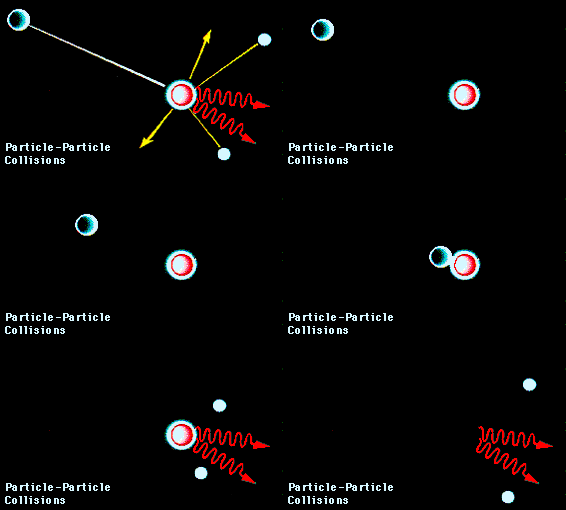
1. X-ray fluorescence - X-rays
are formed by the reaction of fast-coming external electrons with electrons in
a metal atom. The spectrum of X-rays generated by this technology varies
according to the metal used.

X-rays require tens of thousands
of volts of voltage. Radiation isotopes help to obtain X-rays without
electricity in places where there is no electricity. X-rays appear when
energetic electrons collide with a tungsten target in an X-ray tube. At very
high voltages they receive more energy. Such energetic electrons can also be
obtained from isotopes. Strontium emits 90 β particles (electrons). Strontium
90 is taken in an insulated lead. The electrons emitted from it are set to hit
the tungsten target. The concentration of X-rays emitted in this method is low.
The tool is set up safely. In 2010, X-rays were used to diagnose and
treat 5 billion medical morphology studies worldwide.
Source By: Wikipedia
Information: Dr. P. Ramesh, Assistant
Professor of Physics, Nehru Memorial College, Puthanampatti, Trichy.

.png)
.jpg)
No comments:
Post a Comment